Thyroid Ultrasonography
PART II : Thyroid Ultrasonography
STEP 1 : The ultrasound probe should be placed transversely on the anterior neck at the level of the isthmus. It is recommended to scan superiorly to the hyoid bone to detect the rare possibility of a thyroglossal cyst. The sonographer should then scan inferiorly to the upper mediastinum. Visualization of various structures including the thyroid lobes, vocal cords, and level 6 lymph nodes (central neck lymph nodes) will be required during this initial step.
STEP 2 : Swing the probe to the right side of the midline to visualize the right thyroid lobe. Scan in the transverse view and visualize this lobe from it’s superior through to it’s inferior pole. Rotate the probe 90degrees clockwise to acquire views of the right thyroid gland in the sagittal plane (from it’s medial border close to the trachea through to it’s lateral border, close to the internal carotid artery). Repeat these exact steps for the opposite left thyroid lobe.
Fig 1.0 A video showing the isthmus, thyroid lobes and trachea in the anthropomorphic neck of a phantom.
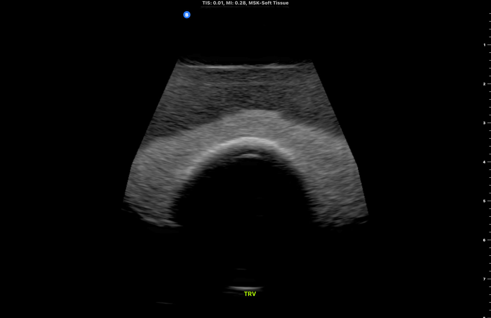
Fig 2.0 Transverse view (TRV) of the thyroid gland
STEP 3 : Return to the thyroid isthmus and measure its size.
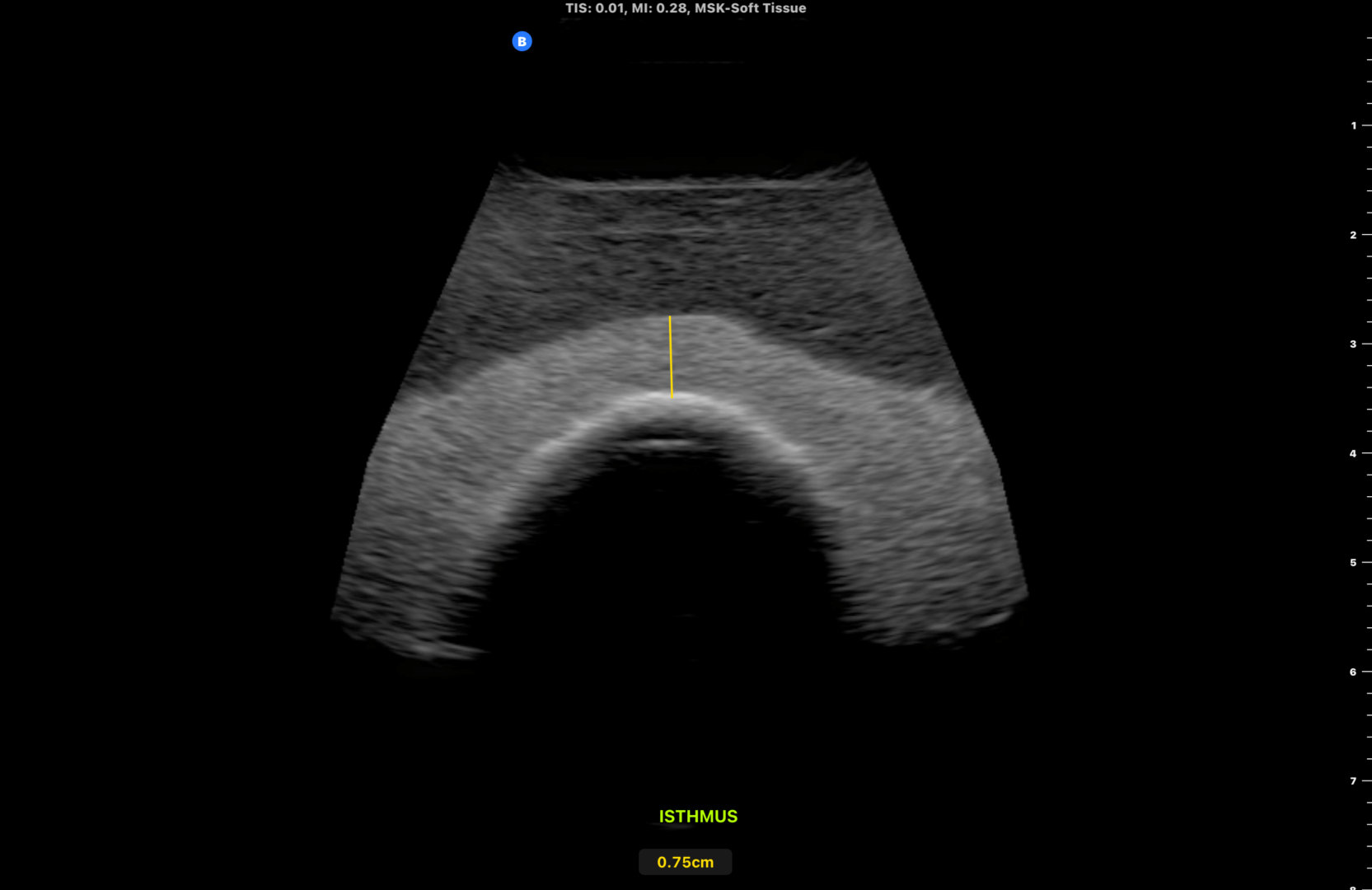
Fig 3.0 Transverse view of the thyroid isthmus. Measurement of the size of the thyroid isthmus in it’s anteroposterior (AP) axis. The normal thyroid isthmus should be less than 0.5cm.
STEP 4 : Scan the right thyroid lobe in the transverse plane. Obtain the following images – inferior(INF), midpoint (MID)and superior(SUP) views
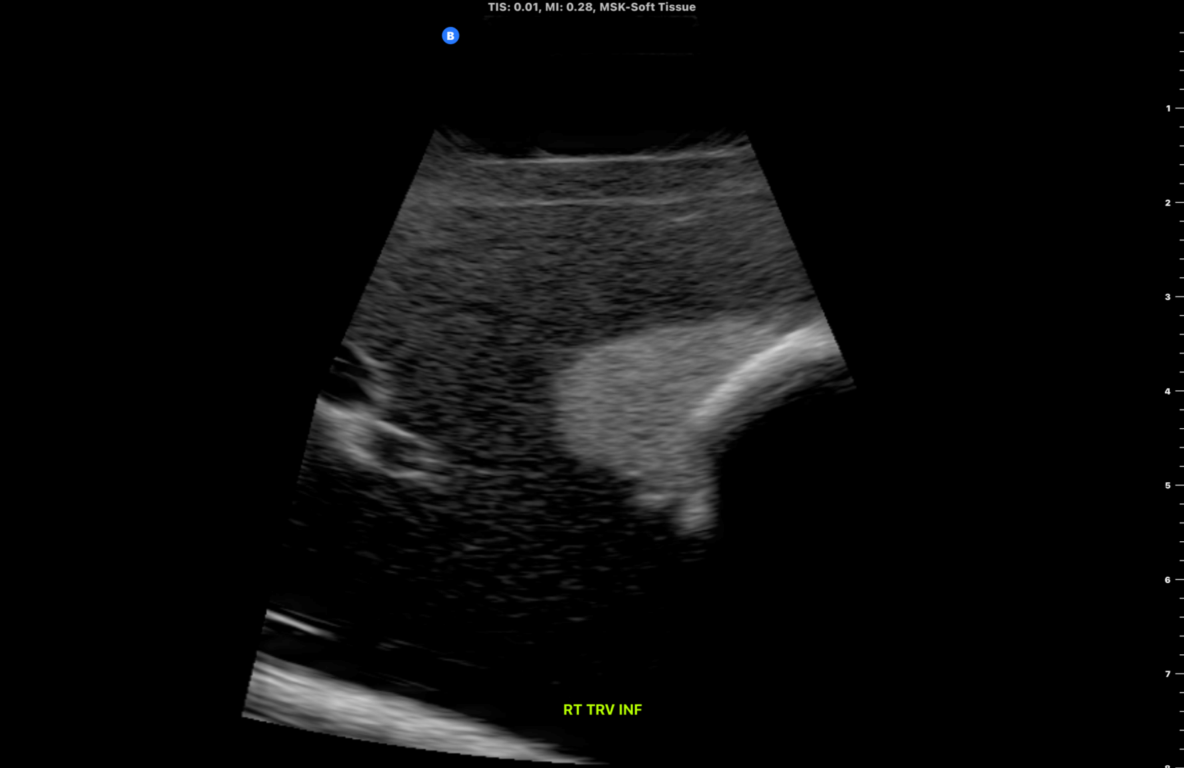
Fig 4.0 Inferior pole of the right thyroid lobe.
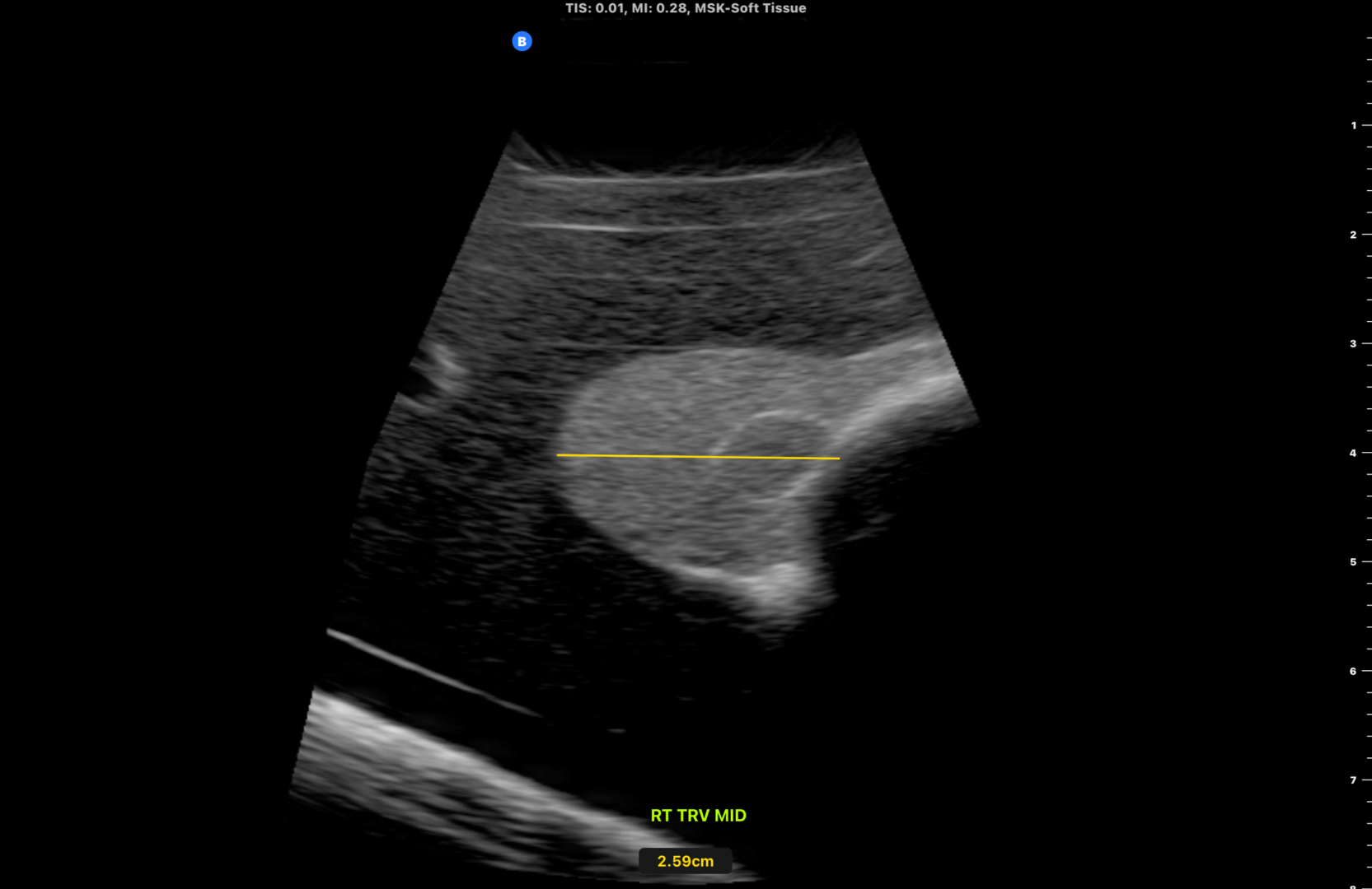
Fig 5.0 Midpoint of the right thyroid lobe showing a subcentimeter isoechoic nodule. Measure the width of the right thyroid lobe. Also measure its size in the AP axis (not shown).
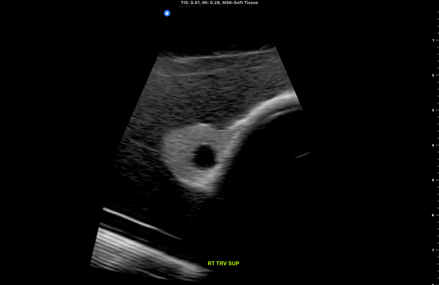
Fig 6.0 Superior pole of the right thyroid lobe.
STEP 5 : Perform a video sweep of the right thyroid lobe from it’s inferior to superior pole (transverse plane)
Fig 7.0 Scanning the right thyroid lobe from it’s inferior to superior poles (Video)
STEP 6 : Scan the right thyroid lobe in the sagittal (longitudinal) plane. Obtain the following images – lateral(LAT), midpoint (MID)and medial (MED) views
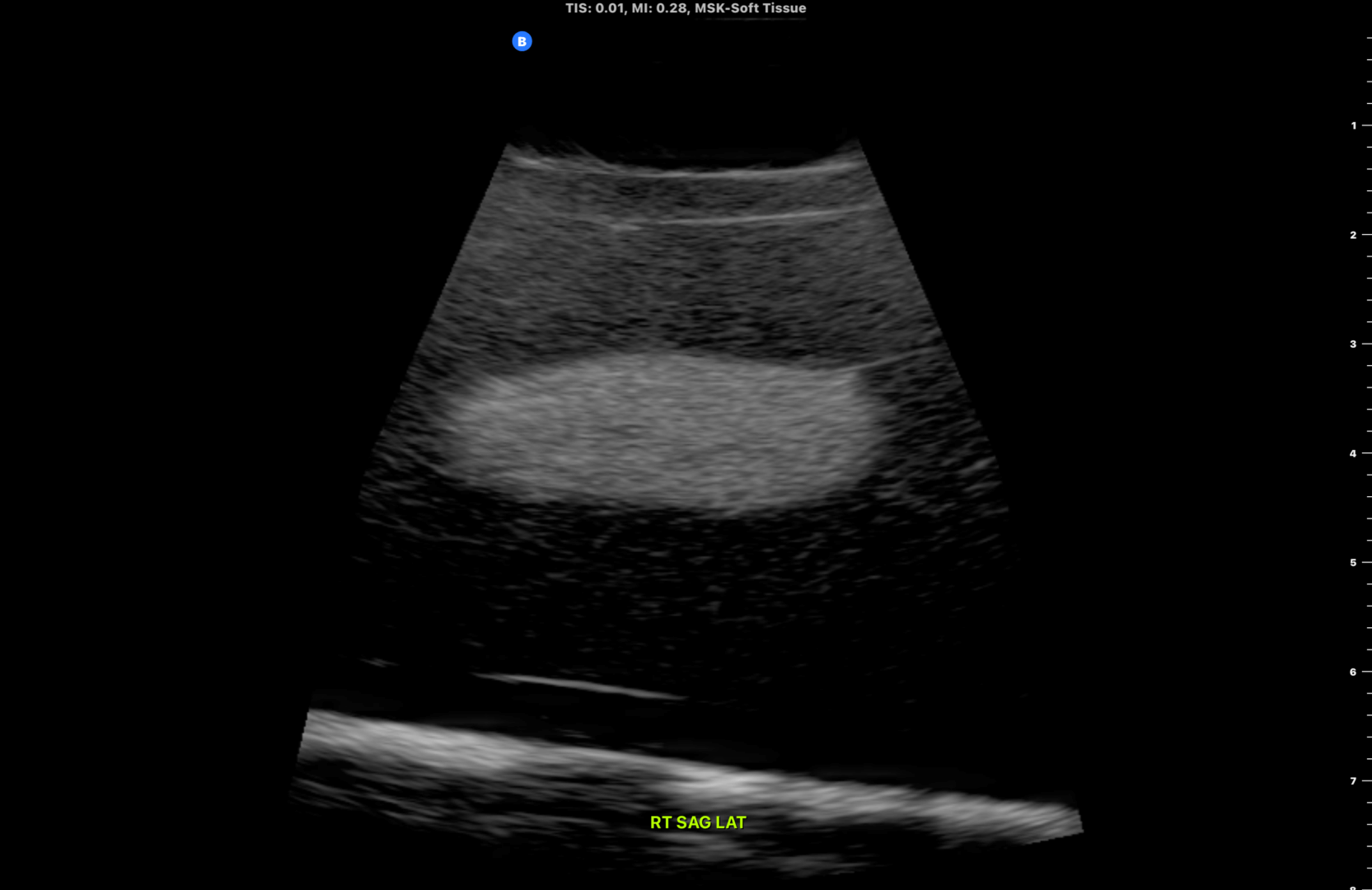
Fig 8.0 Lateral aspect of the right thyroid lobe in the longitudinal plane (close to the internal carotid artery)
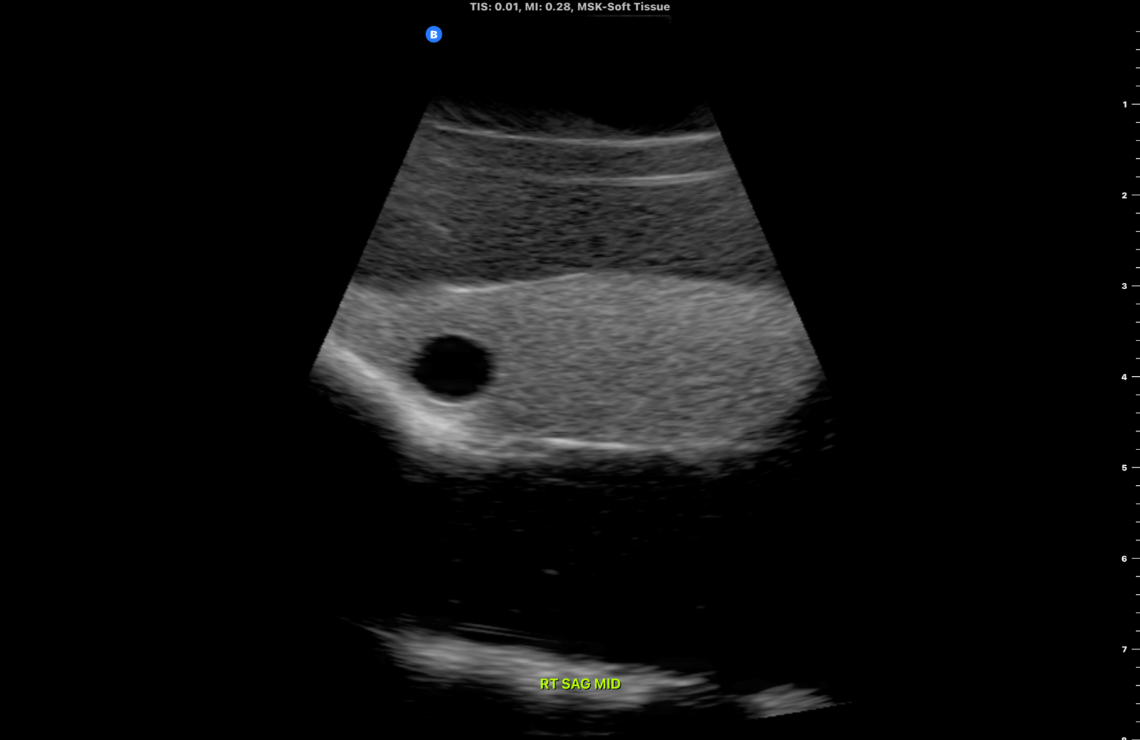
Fig 9.0 Midpoint of the right thyroid lobe in the longitudinal plane. Subcentimeter cystic nodule noted close to the superior aspect of the right lobe. Measure the length of the thyroid lobe from it’s superior to inferior lobe.
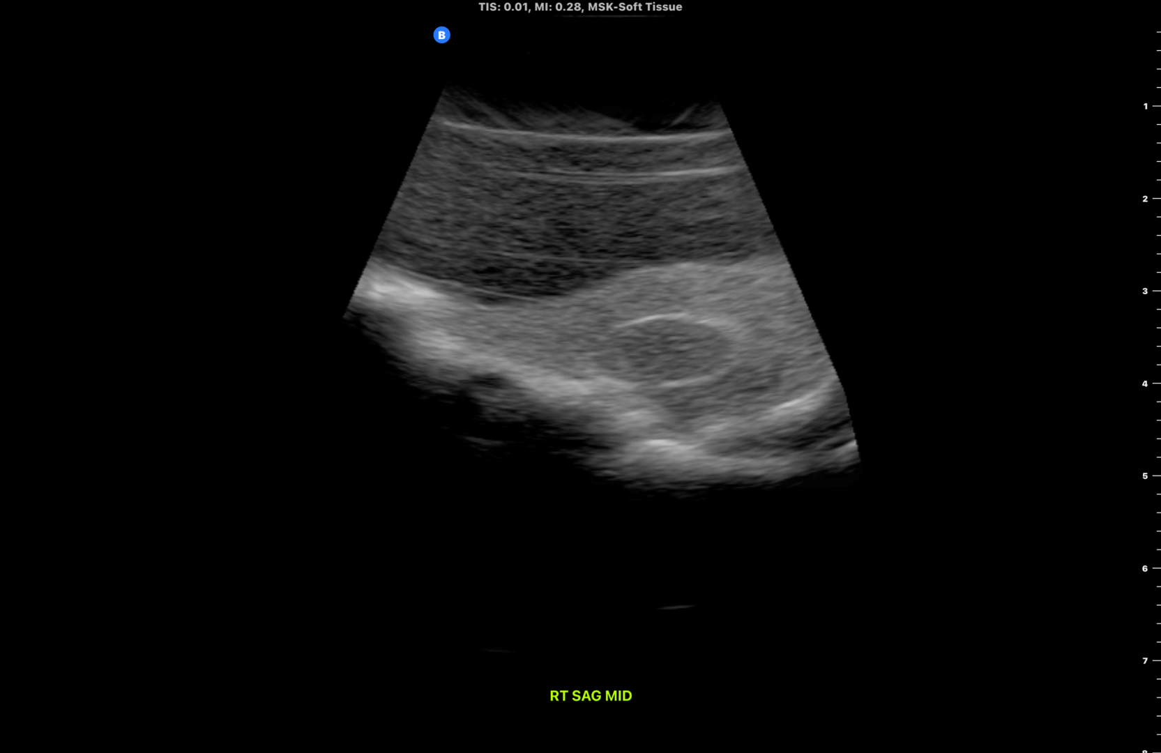
Fig 10.0 Medial aspect of the right thyroid lobe in the longitudinal plane (close to the trachea)
STEP 7 : Perform a video sweep of the right thyroid lobe from it’s lateral to medial aspect (longitudinal plane)
Fig 11.0 Scanning the right thyroid lobe from it’s lateral to medial aspect (Video)
STEP 8 : Repeat the above steps for the left thyroid lobe
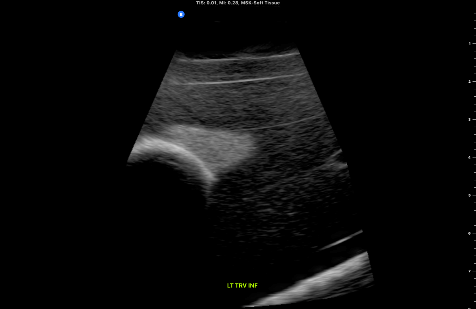
Fig 12.0 Inferior aspect of the left thyroid lobe
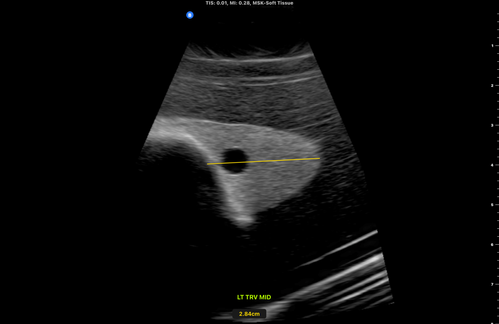
Fig 13.0 Midpoint of the left thyroid lobe. Measure its size in the transverse plane. A cystic subcentimeter nodule is shown.
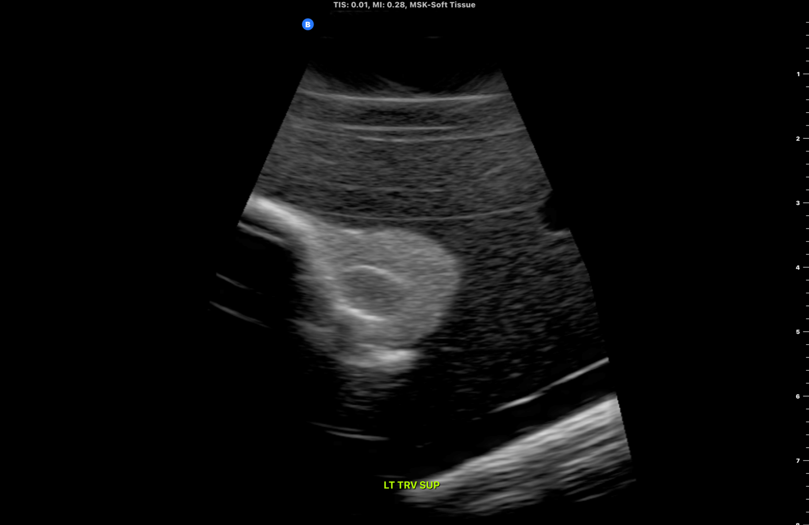
Fig 14.0 Superior aspect of the left thyroid lobe.
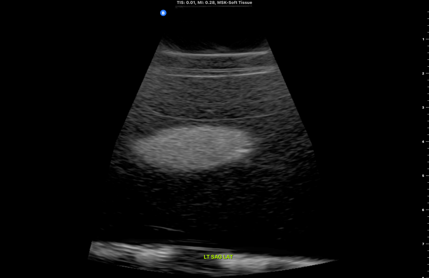
Fig 15.0 Lateral aspect of the left thyroid lobe in the longitudinal plane (close to the internal carotid artery)
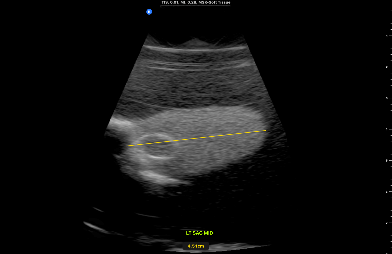
Fig 16.0 Midpoint of the left thyroid lobe in the longitudinal plane. Subcentimeter isoechoic nodule noted close to the superior aspect of the left lobe. Measure the length of the thyroid lobe from it’s superior to inferior lobe.

Fig 17.0 Medial aspect of the left thyroid lobe in the longitudinal plane (close to the trachea)
Perform a video sweep of the left thyroid lobe from it’s lateral to medial aspect (longitudinal plane)
Fig 18.0 Scanning the left thyroid lobe from it’s lateral to medial aspect (Video)
Finally perform a sweep of the right and left lymphatic chains (inferior to superior)