

The islets cells of the pancreas (also known as islets of Langerhans) are somewhat ovoid groupings of pancreatic endocrine cells embedded within a rich background of acinar exocrine tissue. Endocrine cells of the pancreas represent about 2% of the pancreatic volume, with most of the islets being present in the tail of the pancreas. The islets of Langerhans in the pancreas are composed of various cells, including beta, alpha, delta, PP, epsilon, G, and EC cells.
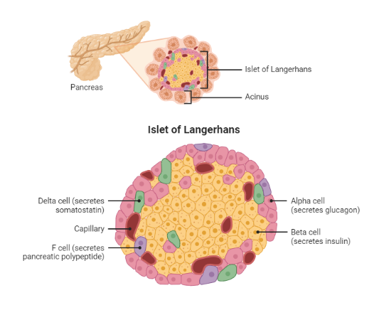
Location and distribution of islet cells of the pancreas.
A normal adult pancreas is composed of approximately one million islet cells. The beta cells are polyhedral in shape and evenly dispersed throughout the pancreas. Alpha cells are columnar in shape and are present principally in the body and tail of the pancreas. The delta cells have a dendritic and are variably distributed. The PP cells are present in the head and uncinate process of the pancreas.
Table 1. Pancreatic islet cells and their corresponding hormones
| Islet cell (frequency) | Hormone produced | Effects(s) |
|---|---|---|
| Beta cells (50-70%) | Insulin and amylin | Insulin increases peripheral glucose uptake and reduces hepatic gluconeogenesis and glycogenolysis. Amylin slows gastric emptying and stimulates satiety |
| Alpha cells (20-30%) | Glucagon | Stimulates hepatic gluconeogenesis and glycogenolysis. Stimulates hepatic ketogenesis during a prolonged fast |
| Delta cells (10%) | Somatostatin | Inhibits the secretion of insulin, glucagon, and PP |
| PP cells (2%) | Pancreatic polypeptide | Inhibition of glucagon secretion and acts as a satiety hormone |
| Epsilon cells (1%) | Ghrelin | Inhibits insulin release after a glucose load. Stimulates GH secretion and is also called the "hunger hormone." |
| G cells (absent) | Gastrin | Pancreatic gastrin-producing cells are present during embryonic development but undergo involution in adults. Re-expression of gastrin can, however, occur in the setting of pancreatic neuroendocrine tumorigenesis (Zollinger-Ellison Syndrome). |
| EC cells (rare) | Serotonin | Classic features of carcinoid syndrome |
GH growth hormone, EC enterochromaffin cells, PP Pancreatic polypeptide.
Histology of the islet cells of Langerhans
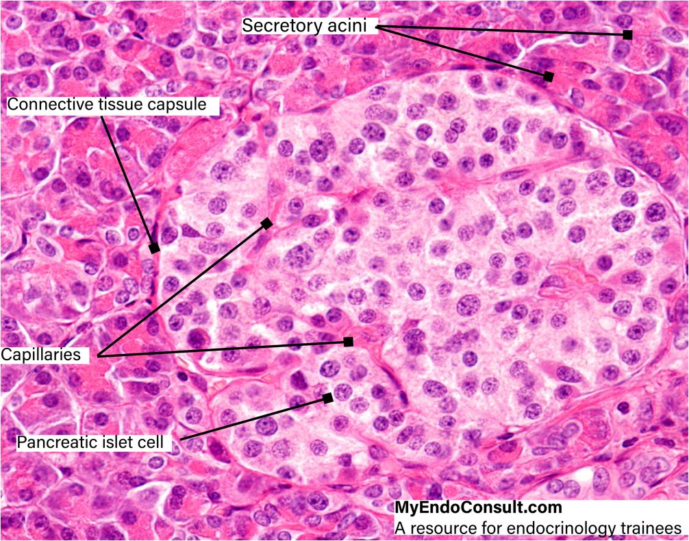 Figure 2. A high-resolution photomicrograph of a pancreas islet (Islet of Langerhans). Stain: Hematoxylin and Eosin
The endocrine pancreatic islet appears as a pale-staining complex in the middle of the field. It lies on a background of deeply staining pancreatic secretory acini (exocrine pancreas). Also, the pancreatic islet contains a rich vascular supply (capillaries) and multiple islet cells encased in a thin connective tissue capsule.
Clinical Pearl (Functional Anatomy of Endocrine Glands)
Pancreatic neuroendocrine tumors (NETs) are derived from pancreatic ductal stem cells that differentiate into the various pancreatic cell subtypes
Table 2. Neuroendocrine tumors of the pancreas
| Neuroendocrine tumor | Clinical feature(s) (pathophysiology) |
|---|---|
| Insulinoma | • Whipple's triad (hyperinsulinemia-induced reduction in glycogenolysis and gluconeogenesis. Also, insulin promotes increased glucose uptake in peripheral tissues) • Weight gain (anabolic effects of insulin) |
| Gastrinoma | • Multiple peptic ulcers (hypergastrinemia mediated increased gastric acid output) • Secretory diarrhea (increased gastric acid output reduces bowel transit time) |
| PPoma (pancreatic polypeptide tumor) | • Weight loss and abdominal discomfort (stimulation of gastrointestinal enzyme secretions) |
| Somatostatinoma | • Complicated gallstones (somatostatin reduces gallbladder contractility and secretions leading to sludging and stone formation) • Steatorrhea (impaired release of bilious fluid from the gallbladder) • Hyperglycemia complications (inhibition of insulin secretion) |
References
- Da Silva Xavier G. The Cells of the Islets of Langerhans. J Clin Med. 2018 Mar 12;7(3):54.
- In’t Veld P, Marichal M. Microscopic anatomy of the human islet of Langerhans. Adv Exp Med Biol. 2010;654:1-19.
Kindly Let Us Know If This Was helpful? Thank You!